Hassanain Qambari and Jayden Dickson
Millions of Americans with diabetes (about 1 in 5) face the risk of eventual blindness due to diabetic retinopathy, a condition that affects blood vessels in the retina, the light-sensitive tissue in the back of the eye. It’s a difficult condition to spot in its earliest stages, since many people don’t show immediate symptoms (although one 2021 study identified key biomarkers that potentially could one day help with early identification). By the late stages, the damage is often irreversible.
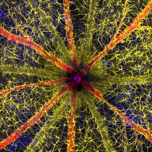
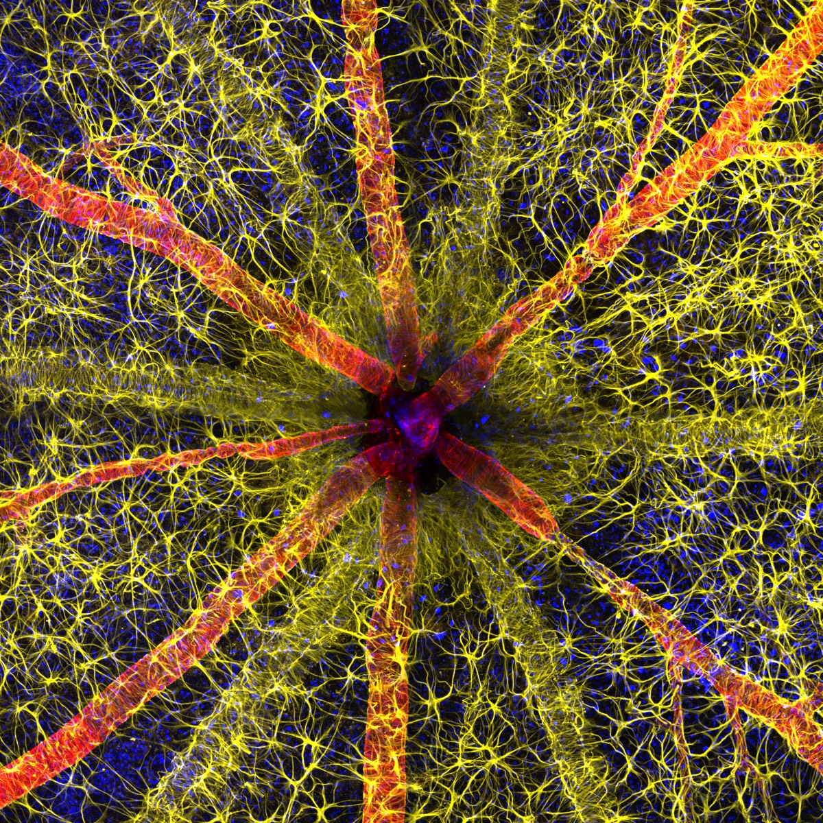
Hassanain Qambari’s research at the Lions Eye Institute in Perth, Australia, focuses on early detection and possible reversal of diabetic retinopathy, including taking precise images of the tiny micron-sized vessels in the eye. With colleague Jayden Dickson’s assistance, he created the winning image in the 2023 Nikon Small World Photomicrography Competition, depicting an optic nerve head in a rodent in exquisite detail.
Now in its 49th year, the annual competition is designed to highlight “stunning imagery from scientists, artists, and photomicrographers of all experiences and backgrounds from across the globe,” according to Nikon’s communications manager, Eric Flem, adding, “I am consistently awed by how these advancements make it possible to create art out of science for the public to enjoy.” Photomicrography involves attaching a camera to a microscope (either an optical microscope or an electron microscope) so that the user can take photographs of objects at very high resolutions. British physiologist Richard Hill Norris was one of the first to use it for his studies of blood cells in 1850, and the method has increasingly been highlighted as art since the 1970s.
Qambari had to overcome a few technical challenges to capture his winning image, not just achieving sufficiently high resolution to showcase fine vessels but also developing a protocol for color-coding the different cell types. Astrocytes are shown in yellow, contractile proteins in red, and retinal vasculature in green.
“The visual system is a complex and highly specialized organ, with even relatively minor perturbations to the retinal circulation are able to cause devastating vision loss,” said Qambari. “I entered the competition as a way to showcase the complexity of retinal microcirculation. Such a competition not only celebrates the participant’s hard work and passion but may also draw and inspire young scientists to pursue a career in STEM. It certainly inspired me.”
Here are the remaining top 20 winners of this year’s contest, ranging from a stunning image of a matchstick igniting and an extreme closeup of tarantula fangs, to micro-peeks at crystallized sugar syrup and the wing scales of a Chinese moon moth. You can check out the full list of winners, as well as several honorable mentions, here.
-
Second place: a matchstick igniting by the friction surface of a matchbox.
Ole Bielfeldt -
Third place: breast cancer cells.
Malgorzata Lisowska -
Venomous fangs of a small tarantula.
John-Oliver Dum -
Auto-fluorescing defensive hairs covering the leaf surface of Eleagnus augustfolia exposed to UV light.
David Maitland -
Slime mold showing capillitial fibers through its translucent peridium.
Timothy Boomer -
Mouse embryo.
Grigorii Timin and Michael Milinkovitch -
Caffeine crystals.
Stefan Eberhard -
Cytoskeleton of a dividing myoblast: tubulin (cyan), F-actin (orange), and nucleus (magenta).
Vaibhav Deshmukh -
Motor neurons grown in microfluidic device for separation of cell bodies (top) and axons (bottom). Microtubules are in green, growth cones are in red.
Melinda Beccari and Don W. Cleveland -
Crystallized sugar syrup.
Diego Garcia -
“Cuckoo wasp” standing on a flower.
Sherif Abdallah Ahmed -
Blood and lymphatic vasculatures in the ear skin of an adult mouse
Satu Paavonsalo and Sinem Karaman -
Sunflower pollen on an acupuncture needle.
John-Oliver Dum -
Fluorescent image of an Acropora sp. showing individual polyps with symbiotic zooxanthellae.
Pichaya Lertvilai -
Carbon nanotubes.
Diego Garcia -
Chinese moon moth wing scales.
Yuan Ji -
A cryptocrystalline micrometeorite resting on a testing sieve.
Scott Peterson -
Stomata in peace lily leaf epidermis.
Marek Miś -
Adult transgenic zebrafish head showing blood vessels (blue), lymphatic vessels (yellow), and the skin and scales (magenta).
Daniel Castranova and Brant Weinstein

